Seminar 1: How to achieve super-resolution imaging in ultrasound in 2D and 3D? A broad assessment of single particle localization algorithms in ULM and of available techniques for super-resolution
Date: 27 August 2019 (Tuesday), 10:00 AM
Venue: 新金博大厦 208 室
Invited Speakers: Baptiste Heiles
报告人:Baptiste Heiles博士,法国Langevin研究所
Abstract: Ultrasound localization microscopy (ULM) has been demonstrated to overcome the penetration/resolution paradigm [Couture et al, review IEEE-TUFFC, 2018]. It relies on localizing the centre of a single microbubble and accumulating a large number of echoes to produce micrometric images of in-vitro canals in 2D [Couture et al. IUS, 2011, Viessman et al, PMB, 2013] and in 3D [Desailly et al, APL, 2013, Heiles et al, IEEE-TMI 2019] and in-vivo, rat brain microvasculature [Errico et al, Nature, 2015, Heiles et al IEEE-IUS 2018], tumours [Lin et al, Theranostics, 2017]. In this presentation we will present a state of the art of different ULM techniques in both 2D and 3D and of other techniques achieving super-resolution imaging and their use as a diagnostics tool. We will also present an overview of the different algorithms used to perform single particle localization for super-resolution image formation using contrast agents and a comparison of their main pros and cons. To demonstrate these, we will use the latest 2D and 3D in vivo imaging of organs in rodents.
Bio-sketch: Baptiste Heiles
Baptiste Heiles was born in France in 1990. He received a Master of Science in General and Civil Engineering from Ecole Normale Superieure Paris Saclay in 2013, the agrégation de Sciences Industrielles in 2014, and a Master of Research in Fluid Mechanics and Heat Transfers from Ecole Centrale de Paris in 2015. Following the work of Yann Desailly and Claudia Errico, he works on exploiting contrast agents for imaging. He focuses on implementing Ultrasound Localization Microscopy (uULM) in volumetric imaging as well as improving ULM in conventional 2D imaging.

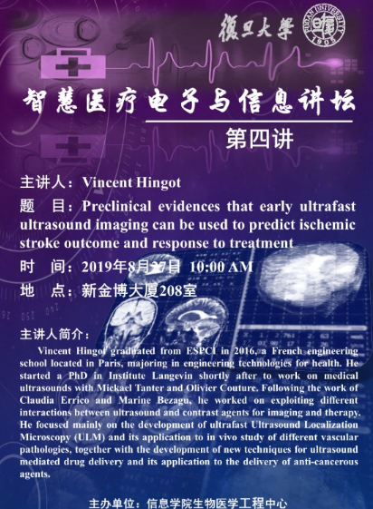
Seminar 2: Preclinical evidences that early ultrafast ultrasound imaging can be used to predict ischemic stroke outcome and response to treatment.
Date: 27 August 2019 (Tuesday), 10:00 AM
Venue: 新金博大厦 208 室
Invited Speakers: Vincent Hingot
报告人:Vincent Hingot博士,法国Langevin研究所
Abstract: In the field of ischemic cerebral injury, a consensus is that tissue perfusion is associated with a favorable clinical outcome. Thus, ideal patient selection for current treatments should not be based on therapeutic windows but rather on perfusion imaging in order to determine “a signature” of response to treatments. It is thus admitted that advanced imaging-based approaches able to better diagnose and prognose those patients would contribute to improve the clinical care. However, current imaging modalities do not yet provide such advanced diagnosis. Ultrafast Ultrasound is a powerful tool to perform long term follow-up of the cerebral blood flow, non-invasively, with high spatial and temporal resolution and high sensitivity. In this study, in a model of thromboembolic stroke in mice, subjected to the gold standard pharmacological stroke therapy, we provide the proofs of concept that Ultrasound Localization Microscopy, performed very early after stroke onset and through the skull is predictive of final outcome and can be used to early unmask response to treatment. This innovative study thus provides in addition of new insights on tissue perfusion and suffering following ischemic stroke, the information needed for a future translation of Ultrafast Ultrasound Imaging to the clinical setting in the context of stroke.
Bio-sketch: Vincent Hingot
Vincent Hingot graduated from ESPCI in 2016, a French engineering school located in Paris, majoring in engineering technologies for health. He started a PhD in Institute Langevin shortly after to work on medical ultrasounds with Mickael Tanter and Olivier Couture. Following the work of Claudia Errico and Marine Bezagu, he worked on exploiting different interactions between ultrasound and contrast agents for imaging and therapy. He focused mainly on the development of ultrafast Ultrasound Localization Microscopy (ULM) and its application to in vivo study of different vascular pathologies, together with the development of new techniques for ultrasound mediated drug delivery and its application to the delivery of anti-cancerous agents.


